ECG
Electrocardiography, or ECG, is a crucial diagnostic tool that is widely used in the field of cardiology. At CoreMed Plus, we understand the importance of early detection and accurate diagnosis of heart conditions, and ECG testing plays a significant role in achieving these goals. Our team of skilled healthcare professionals is dedicated to providing our patients with the highest level of care using state-of-the-art technology and evidence-based practices.
At CoreMed Plus, we offer a range of ECG testing services, including routine ECGs, exercise stress tests, and Holter monitoring. Routine ECGs are typically performed in our office and take only a few minutes to complete. Exercise stress tests involve monitoring the patient’s heart activity while they walk on a treadmill or ride a stationary bike. Holter monitoring involves wearing a small device that records the patient’s heart activity throughout 24 to 48 hours.
Our team of healthcare professionals is committed to providing our patients with the highest level of care using the latest technology and evidence-based practices. We understand that heart conditions can be stressful and overwhelming, and we strive to provide our patients with a supportive and compassionate environment where they can feel comfortable discussing their concerns and receiving the care they need.
In addition to our ECG testing services, we offer various other cardiology services, including echocardiography, stress echocardiography, and vascular testing. Our team of healthcare professionals includes board-certified cardiologists, nurses, and other specialists who work together to provide comprehensive care to our patients.
At CoreMed Plus, we are committed to providing our patients with the highest level of care using the latest technology and evidence-based practices. Our goal is to help our patients achieve optimal heart health and improve their quality of life. Every patient deserves personalized care tailored to their needs, and we work closely with each patient to develop a customized treatment plan for them.
Electrocardiography is a crucial diagnostic tool that is widely used in the field of cardiology. At CoreMed Plus, we offer a range of ECG testing services to help diagnose and manage heart conditions. Our team of healthcare professionals is committed to providing our patients with the highest level of care using the latest technology and evidence-based practices. Every patient deserves personalized care tailored to their needs, and we strive to create a supportive and compassionate environment where our patients can feel comfortable discussing their concerns and receiving the care they need.

What is Electrocardiography
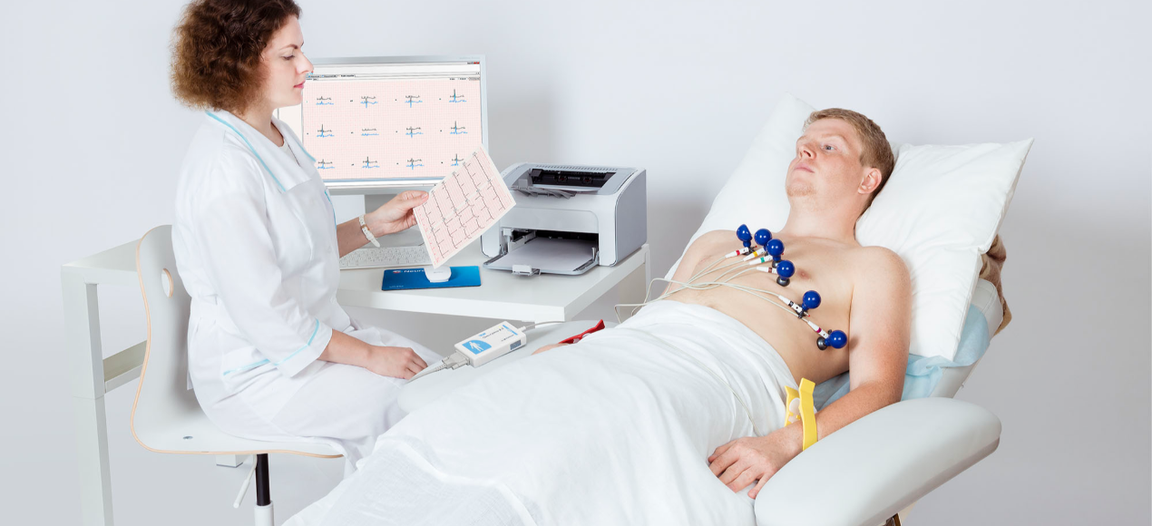
Electrocardiography, or ECG, is a medical test commonly used to assess the heart’s electrical activity. An ECG test involves placing electrodes on the skin of the chest, arms, and legs, which are then connected to a machine that records the electrical signals generated by the heart. These signals are represented on a graph called an electrocardiogram.
The heart is a muscular organ that pumps blood throughout the body. It is made up of four chambers: the right and left atria and the right and left ventricles. The atria are the heart’s upper chambers and receive blood from the body or lungs, while the ventricles are the lower chambers of the heart and pump blood out to the body or lungs.
The heart’s electrical system controls the heart’s rhythm and ensures that the atria and ventricles contract in a coordinated manner. A specialized group of cells generates the electrical signals that control the heart’s rhythm called the sinoatrial (SA) node. The SA node is located in the right atrium. It acts as the heart’s natural pacemaker, generating electrical impulses that travel through the atria and ventricles, causing them to contract and pump blood.
An ECG test measures the heart’s electrical signals as it contracts and relaxes. The test is non-invasive and painless and usually takes only a few minutes to complete. The patient remains on a table during the test while the electrodes are attached to their skin. The electrodes are connected to a machine that records the electrical signals generated by the heart.
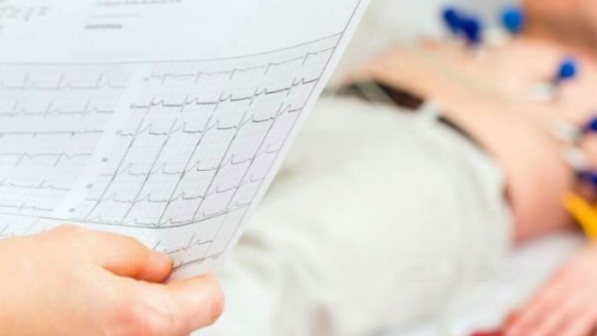
The resulting electrocardiogram shows the heart’s electrical activity as a series of waves. These waves represent the electrical signals generated as the heart contracts and relaxes. Each wave has a specific name and illustrates a particular event in the cardiac cycle.
The P wave represents the electrical activity of the atria as they contract and pump blood into the ventricles. The QRS complex represents the electrical activity of the ventricles as they contract and pump blood out of the heart. The T wave represents the electrical activity of the ventricles as they relax and prepare for the next contraction.
An ECG test can be used to diagnose a variety of heart conditions, including arrhythmias, heart attacks, and heart failure. Arrhythmias are abnormal heart rhythms that can be caused by a variety of factors, including heart disease, stress, and certain medications. A heart attack occurs when the blood flow to the heart is blocked, causing damage to the heart muscle. Heart failure occurs when the heart cannot pump enough blood to meet the body’s needs.
ECG tests are also used to monitor the effectiveness of certain heart treatments, such as pacemakers and anti-arrhythmic medications. A pacemaker is a small device implanted under the chest’s skin and connected to the heart with wires. The pacemaker sends electrical signals to the heart, helping it maintain a regular rhythm. Anti-arrhythmic medications are drugs that help control abnormal heart rhythms.
In addition to its diagnostic and monitoring uses, ECG testing is also used in research to study the heart’s electrical activity and to develop new treatments for heart disease. Researchers use ECG testing to analyze the effects of drugs and other medicines on the heart’s electrical activity and identify new therapeutic targets.
Electrocardiography is an essential tool in the diagnosis and management of heart disease. By measuring the heart’s electrical activity, ECG testing can provide valuable information about the heart’s function and help healthcare providers make informed treatment decisions.
Why an Electrocardiography May be Required
Electrocardiography, also known as an ECG or EKG, is a non-invasive test that records the heart’s electrical activity. An ECG can provide valuable information about the heart’s rhythm, rate, and overall function. There are several reasons why someone may require an ECG, including:
- To Diagnose a Heart Condition: An ECG can help diagnose a wide range of heart conditions, such as arrhythmias, coronary artery disease, heart attacks, and heart failure. The test can also help identify structural abnormalities in the heart, such as an enlarged heart or a valve disorder.
- To Assess Heart Function: An ECG can provide information about how well the heart functions. The test can help determine if the heart is receiving enough oxygen and if the heart’s chambers are contracting and relaxing properly.
- To Monitor Treatment: If someone has a known heart condition, an ECG may be used to monitor their response to treatment. For example, an ECG may be used to assess medication effectiveness or determine if a surgical procedure was successful.
- To Evaluate Symptoms: If someone is experiencing symptoms such as chest pain, shortness of breath, or palpitations, an ECG may be ordered to help determine the cause of the symptoms. The test can help rule out or diagnose heart-related conditions contributing to the symptoms.
- As Part of a Routine Physical Exam: An ECG may be included as part of a routine physical exam, especially for individuals who have risk factors for heart disease, such as high blood pressure, diabetes, or a family history of heart disease. This can help detect heart problems early on and prevent more severe complications from developing.
An ECG is a valuable tool that can provide important information about the heart’s function and help diagnose various heart-related conditions. If someone is experiencing symptoms or has risk factors for heart disease, their healthcare provider may recommend an ECG as part of their evaluation.

Steps for a Standard ECG Test
- The patient will be asked to remove any jewelry or clothing that might interfere with the placement of the electrodes.
- The patient will be asked to lie still on a table or bed.
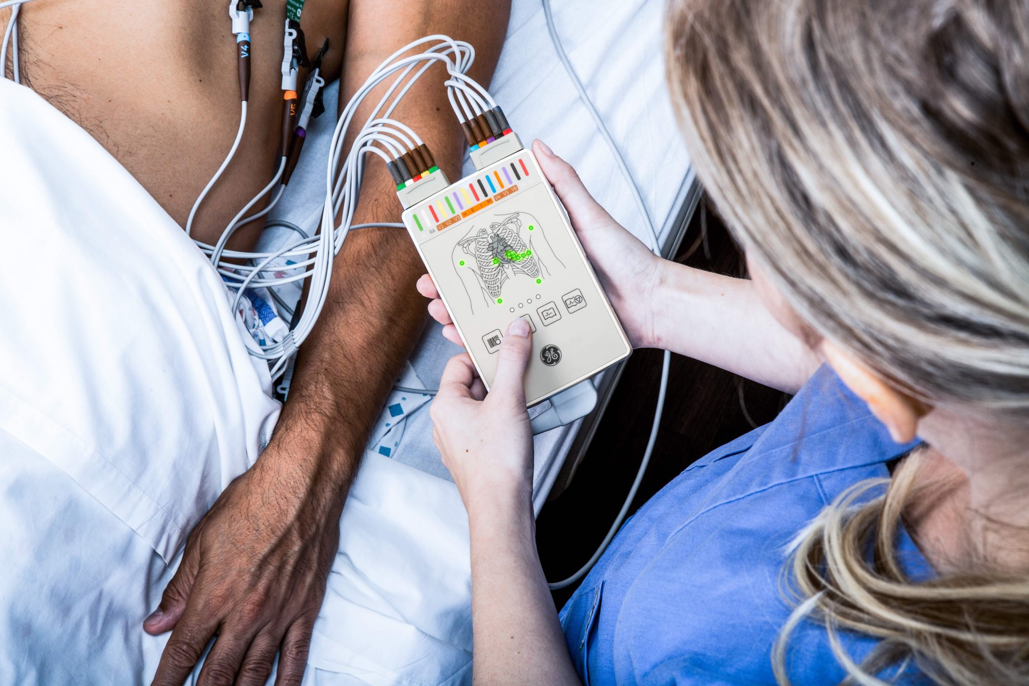
- Electrodes will be placed on the patient’s chest, arms, and legs using a sticky adhesive.
- The electrodes will be connected to a machine that records the heart’s electrical activity.
- The patient may be asked to hold their breath or remain still for a few seconds during the test to ensure an accurate reading.
- Once the test is complete, the electrodes will be removed, and the patient can return to normal activities.
- A healthcare professional will review the test results and provide a diagnosis and treatment plan if necessary.
Note: In some cases, the patient may be asked to perform an exercise stress test involving walking on a treadmill or riding a stationary bike while the heart is monitored with ECG electrodes. The steps for this type of test may vary slightly from a routine ECG test.
Why Choose CoreMed Plus
Here are some reasons why you should choose CoreMed Plus for your ECG needs:
- Experienced Healthcare Professionals: Our team of board-certified cardiologists, nurses, and other specialists have years of experience in cardiology and are dedicated to providing our patients with the highest level of care.
- State-of-the-Art Technology: We use the latest technology to provide accurate and reliable ECG testing results, including routine ECGs, exercise stress tests, and Holter monitoring.
- Comprehensive Care: Besides ECG testing, we offer a range of other cardiology services, including echocardiography, stress echocardiography, and vascular testing.
- Personalized Care: Every patient deserves customized care tailored to their needs. We work closely with each patient to develop a customized treatment plan.
- Compassionate Environment: We understand that heart conditions can be stressful and overwhelming. Our team of healthcare professionals provides a supportive and compassionate environment where our patients can feel comfortable discussing their concerns and receiving the care they need.
- Convenient Location: Our office is conveniently located and easily accessible, making it easy for our patients to receive the care they need.
- Efficient Service: We value our patients’ time and strive to provide efficient service without compromising the quality of care.
- Evidence-Based Practices: We use evidence-based practices to guide our treatment decisions, ensuring patients receive the most effective and up-to-date care.
Commitment to Patient Satisfaction: Our goal is to help our patients achieve optimal heart health and improve their quality of life. We are committed to patient satisfaction and go above and beyond to ensure our patients are happy with their care.

Don't Delay Call Today!
CoreMed Plus is committed to providing our patients with the highest level of care using the latest technology and evidence-based practices. We understand that heart conditions can be stressful and overwhelming. Still, early detection and accurate diagnosis are crucial to managing heart disease and improving our patient’s quality of life.
So, if you require ECG testing or other cardiology services, we invite you to contact us today to schedule an appointment. Our experienced team of healthcare professionals is dedicated to providing personalized care that is tailored to your individual needs, and we will work closely with you to develop a customized treatment plan that is right for you.
Take charge of your heart health today and contact CoreMed Plus before it’s too late. We look forward to helping you achieve optimal heart health and quality of life!
Contact Information
If you’re ready to take charge of your health and embark on a journey toward a healthier, happier life, we invite you to contact us today. Our compassionate and knowledgeable staff is excited to take your call and help you on your health journey. We will work closely with you to develop a customized treatment plan that is right for you and provide the support and care you need every step of the way.
Don’t let health concerns hold you back – contact CoreMed Plus today and let us help you achieve optimal health and wellness. We look forward to hearing from you and helping you achieve a healthier, happier life.
- Email: [email protected]
- Phone: +1 248-666-6005


